
Ankle Fractures
Written By: Brian Smith, DO; Edited by: Tim Khowong, MD
Case: A 21-year-old male presents to the Emergency Department for a right ankle injury sustained during a Division I college basketball game.

He has been unable to bear weight on the right leg and has significant tenderness and swelling throughout the ankle on exam.
You obtain Xrays of the ankle, which are shown below:
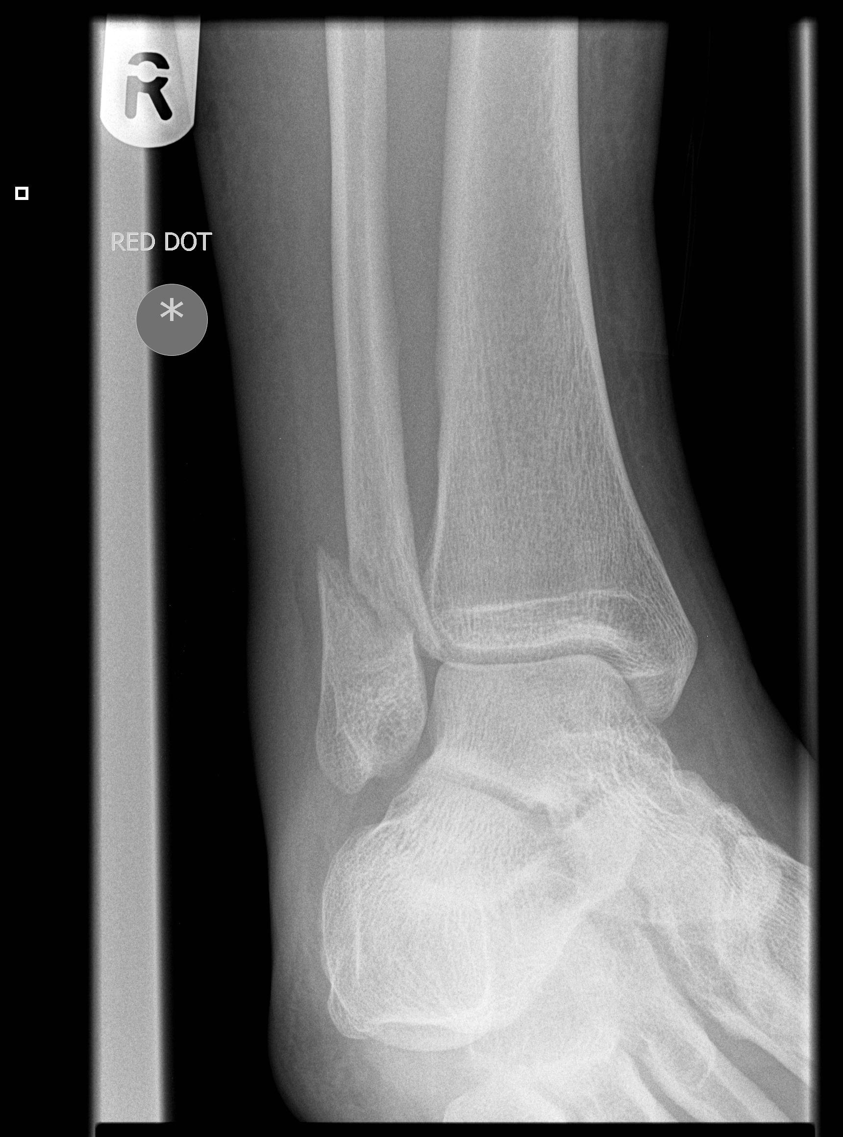

Lateral View
What is the diagnosis? Next Best Step? Management and Disposition?
If you don’t know the answer, that’s OK! By the end of this blog post you’LL be able to:
Classify ankle fractures on Xray
Execute the appropriate management of various ankle fractures
Anatomy:
In order to understand ankle fractures, we first need to know the anatomy:
The ankle joint is composed of 3 malleoli (image 1)
Lateral Malleolus (lateral fibula)
Medial Malleolus (medial tibia)
Posterior Malleolus (posterior tibia)
Additionally, it is important to identify the ankle mortise and area of distal tibiofibular joint (image 2), which is the space between the tibia and talus. This will help us to classify stability of certain ankle fractures. Widening of the ankle mortise is a sign of injury to the deltoid ligament (image 3), which is considered an unstable ankle injury.

Image 1: anatomy of the ankle

Image 2: Distal tibiofibular joint and ankle mortise on x-ray

Image 3: The Deltoid Ligament
Imaging:
There are 3 standard views for ankle xrays:
For AP and Mortise views, it is important to identify:
Medial and lateral malleoli
Overlap between tibia and fibula distally (orange arrow). Normal: >6mm
Ankle mortise medial clear space (blue arrows). Normal: < 4mm
Lateral clear space between fibula and talus (white circle). Normal: 3-6mm
For the Lateral view, it is important to identify:
Posterior malleolus
Distal fibula in relation to tibia. Normal = projection over posterior 1/3 of distal tibia

Image 4: Standard Views on Ankle xrays
Ankle Fractures:
Ankle fractures can be separated into two categories: Stable vs Unstable
Stable ankle fractures can be placed in a short-leg posterior splint and given instructions for weight-bearing, RICE, and follow-up with orthopedics outpatient in 5-7 days
Unstable ankle fractures require orthopedics consult in the Emergency Department for reduction and short-leg posterior splint placement. These fractures require ORIF
But how do we know if an ankle fracture is stable or unstable?
Bimalleolar fractures and Trimalleolar (images 6 & 7) fractures are considered unstable
Isolated Medial malleolar fractures and posterior malleolar fractures are considered stable as long as there is no significant displacement

Image 5: Short-Leg posterior Splint

Images 6 (Left) & 7 (right) : Trimalleolar fracture involving medial and lateral malleoli (left) and posterior malleolus (right)

I know what you’re thinking…What about isolated lateral malleolar fractures???
This is where things get a bit more complicated…
Danis-Weber Classification:
Isolated lateral malleolar fractures are classified by their location on the fibula in relation to the distal tibiofibular (TF) syndesmosis (image 8). Each type has a different management. This can be difficult to keep straight but I’ll make it easy with my mnemonic.

Image 8 (above) = Weber fractures in relation to the TF syndesmosis
Weber A = Avulsion or “A”-OK
Avulsion, fracture distal to TF syndesmosis
Stable
Next Step: Short-leg posterior splint, discharge with orthopedics follow up in 5-7 days
Weber B = Back to x-ray
Fracture at the level of the TF syndesmosis
Can be Stable or Unstable
Next Step: need to obtain stress views (image 10) of ankle to assess for stability
Obtained by using manual stress or gravity on ankle joint
Positive if ankle mortise medial clear space is widened (unstable)
Weber C = Call ortho
Fracture proximal to TF syndesmosis
Unstable
Next Step: Call orthopedics consult for reduction and splinting in ED

Image 9 (left) = Weber A, B, and C fractures seen on x-ray
image 10 (right) = Stress views of Weber B fractures. The xray on the left demonstrates a Weber B fracture with positive stress view, as evident by widening of medial clear space. The rightmost xray shows negative stress views of a Weber B fracture, with no widening of ankle mortise medial clear space; this is a STABLE Weber B fracture.

Back to the Case,
Our patient’s x-rays are shown again below:
Using the tools we just reviewed, you should now be able to:
Classify the fracture pattern seen in imaging
Idenfity the next best step in management

If you said Weber B and obtain stress views, you got it!
But now we still have one more image to interpret. You obtain stress views (pictured below).
Is this stable or unstable?
What is our next step?

Image 11: Stress Views of our patient’s fracture
Based on stress views, this fracture is UNSTABLE, given the widening of the medial clear space.
Orthopedics was consulted and the fracture was reduced and splinted in the ED. This patient then underwent ORIF a few days later.
Take Home Points
Ankle Fractures can be classified as stable or unstable
Stable —> Splint in ED and send home with ortho follow up in 5-7 days
Unstable —> Consult orthopedics for reduction and splinting in ED. Will need ORIF
Bimalleolar and Trimalleolar fractures are unstable
Isolated posterior malleolar and medial malleolar fractures are stable
Isolated lateral malleolar fractures are further categorized via the Weber Criteria
Weber A = Avulsion or “A”-OK - Stable
Weber B = Back to x-ray for stress views
Weber C = Call ortho for immediate reduction and splinting