
Sold by Cold
Brian Smith, DO
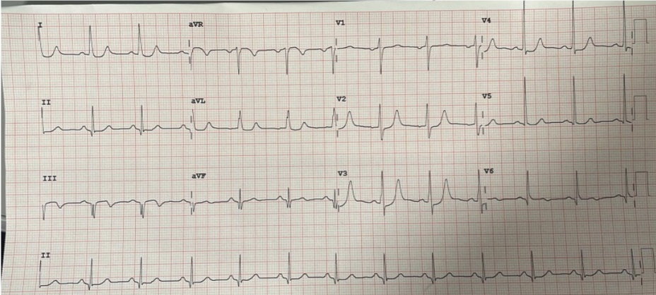
You’re working a busy shift and are handed this This ECG:
You do a quick chart review and see this is a 72 M PMH HTN, HLD, DM, ON ASA 81 who presents with 3 days of epigastric pain that occurs after drinking water. He states pain is not exertional but does improve after taking aspirin. His last episode of epigastric pain was 2 hours prior to arrival, at which time he also had a syncopal episode. No other complaints. He’s currently asymptomatic and appears well. His vital signs are within normal limits.
What do you want to do for this patient?
A) This is concerning for STEMI, activated cath lab
B) Bring the patient to the trauma bay immediately, repeat ECG, obtain serial troponin and imaging
C) Order some labs and send the patient to the main ED with telemetry monitoring
D) Send the patient back to the waiting room
Freeze for a minute before scrolling. What does this ECG pattern represent?
de Winter T Waves:
This is a STEMI Equivalent
Seen in 2% of all LAD occlusions
Diagnostic Criteria:
Tall, prominent, symmetrical T waves in precordial leads (arrows)
Upsloping ST depressions >1mm at point (Asterix)
Can See reciprocal STE in aVR
Cab be preceded or followed by typical STEMI morphology

Remember these "Can’t Miss” STEMI equivalents:

Back To Our Patient:
The patient is comfortable-appearing and resting in stretcher. A bedside ECHO is performed and shows anterior regional wall abnormalities. ECG was repeated, which showed no dynamic changes. STEMI was activated and patient was taken to cath lab. He was found to have a 100% mid-LAD occlusion.
Take Home Point: DeWinter’s Pattern is a STEMI Equivalent
If you see this pattern on ECG, it is a STEMI until proven otherwise
