
Rotator Cuff Ultrasound
Written by: Dr. Faria Rahman
Edited by: Dr. Joann Hsu
Case:
36 year old female presenting with left shoulder pain after trip and fall last night. Left shoulder reduction attempted at urgent care, but patient presents to the ED for persistent left shoulder pain and concern for rotator cuff injury.
Exam:
Patient is in considerable pain, holding her left arm in a flexed position and is unable to move the extremity due to pain.

The head of the humerus is anteriorly displaced from the glenoid fossa, making this an anterior shoulder dislocation.
Ultrasound for shoulder dislocation is a topic in itself. Today, we will focus on rotator cuff injuries on ultrasound!
How to assess for rotator cuff injury?
Although MRI is the gold standard for diagnosing rotator cuff injury, ultrasound is a great bedside tool to use in conjunction with your physical exam in the ED.
We will go through normal anatomy in the below section. Note the parallel, organized appearance of muscle and the organized but more hyperechoic tendons. The parallel fibers in both muscle and tendon should be uninterrupted. Any disruptions or variations from this organized structure should be suspicious for injury (partial or compete tears, etc)
But first, let’s go over the rotator cuff anatomy.
There are four muscles that make up the rotator cuff:
Supraspinatus
Infraspinatus
Subscapularis
Teres Minor.

Supraspinatus: Abducts shoulder
Infraspinatus: Externally rotates shoulder
Teres minor: Externally rotates shoulder
Subscapularis: Internally rotates shoulder
**Teres minor rarely has pathology, so is not usually assessed for injuries**
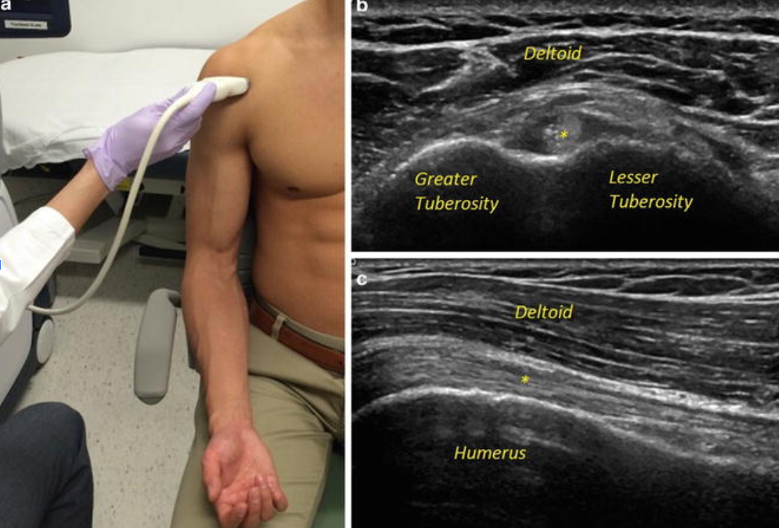
View 1: Biceps Tendon
This view assesses for biceps tendon pathology.
In this first position, you will have the patient resting their arm along their torso with elbow bent 90 degrees and palm facing up, while you scan anterior to the shoulder.
On the right top image, you see the anterior aspect of the shoulder joint in transverse view, with the head of the humerus as the bottom-most hyperechoic line.
Above the humerus you see the supraspinatus muscle and biceps tendon (starred, in cross section) attaching to the humerus, and above that is the deltoid muscle.
In this first view, you will be able to see the long head of the biceps tendon as well as the supraspinatus muscle attaching to the humerus. (As with all ultrasounds, make sure to scan both transverse and sagittally to see the muscles/tendon in both planes!)
View 2: Subscapularis
In the second position, the patient externally rotates their shoulder out to the side, isolating the subscapularis muscle while still scanning anteriorly.

View 3: Supraspinatus
For the third position, have the patient put their arm behind their back while you scan anterior-laterally.
This will isolate the supraspinatus muscle at the greater tuberosity.

View 4: Infraspinatus
The fourth position has the patient touch their contralateral shoulder while you scan the posterior shoulder, isolating the infraspinatus muscle.

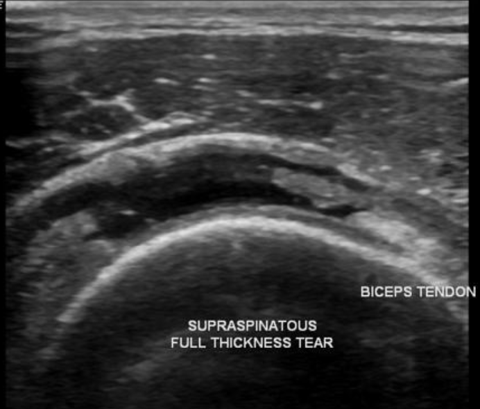
What does a rotator cuff tear look like on ultrasound?
As mentioned earlier, a normal tendon/muscle will look homogenous with striations as you fan up and down along the muscle.
A muscle/tendon tear will have an area of hypoechogenicity that indicates pathology such as a tear or soft tissue edema

Back to our case:
Our patient ended up having just an anterior shoulder dislocation without any obvious signs of rotator cuff injury.
She was asked to follow up in the orthopedic outpatient clinic for reassessment as she was noted to have two consecutive shoulder dislocations within the span of 24 hours.
Happy scanning!
References:
Collin P et al. What is the Best Clinical Test for Assessment of the Teres Minor in Massive Rotator Cuff Tears? Clin Orthop Relat Res. 2015 Sep;473(9):2959-66.
Ultrasound Tutorial: MSK Series: Shoulder / Rotator Cuff | Radiology Nation." Youtube.Com, uploaded by Radiology Nation, 15 Aug. 2020, www.youtube.com/watch?v=BwSJCkTBN0c&t=313s&ab_channel=RadiologyNation.
Moosikasuwan J, Miller T, Burke B. Rotator Cuff Tears: Clinical, Radiographic, and US Findings. Radiographics. 2005;25(6):1591-607. doi:10.1148/rg.256045203 - Pubmed