
Gallbladder Polyps
Written by: Dr. Kyle Soldevilla
Edited by: Dr. Joann Hsu
The case:
A 62 year old male with a history of diabetes and obesity s/p gastric bypass presents to your ED for evaluation of right upper quadrant abdominal pain.
For the past month he’s been having on and off pain particularly worse with meals, however his symptoms acutely worsened today accompanied by nausea and vomiting for about 3 hours.
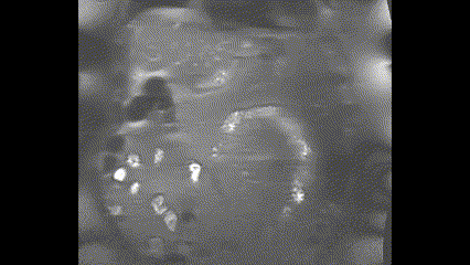
Being the astute Emergency Physician you are, you decide to do a bedside ultrasound which reveals these images:


You think to yourself…what is that abnormal finding seen alongside the stones? Luckily, we’re here to help.
Cholelithiasis vs gallbladder polyps
To start, it is first important to acknowledge that in the image there are two notable findings: cholelithiasis and a gallbladder polyp.
They are easily mistaken for each other but may have different clinical implications!
For the sake of brevity, there are two key differences to help the emergency ultrasonographer distinguish between a polyp and stone.
First, polyps are soft tissue masses and will not cause echogenic shadowing in the same fashion as a stone.
Second, polyps are not mobile. Correspondingly, they should be adherent to the gallbladder wall and should not be mobile if the patient changes positions (i.e. left lateral decubitus positioning).
Now referring back to this clip from the bedside ultrasound:

Here you can clearly see the stones with shadowing along DEPENDENT part of the gallbladder.

Here you can better see the polyp that seems to defy gravity, adherent to the gallbladder wall with NO posterior shadowing.
Gallbladder polyps
Gallbladder polyps are a common cause of biliary colic found in up to 4.5 percent of gallbladders evaluated by ultrasound and 13.8 percent of resected gallbladders.
More often than not, they are a result of gallbladder hyperplasia and lipid deposition as opposed to neoplasia; however imaging alone cannot exclude carcinoma. Histologic examination will be the most definitive testing.
That being said, gallbladder polyps can be classified into two major categories: benign and malignant.
Both benign and malignant lesions share similar ultrasonographic features being echogenic, non-shadowing, and immobile structures, but differ in their underlying etiology
Benign polyps can be further subdivided into non-neoplastic and neoplastic.
In order of frequency, non-neoplastic lesions include: cholesterol polyps, adenomyomas, and inflammatory polyps.
Cholesterol polyps are a characterized by lipid deposition within the gallbladder mucosa.
Adenomyomas are typically a result of mucosal overgrowth, thickening, and intramural diverticula.
Inflammatory polyps are a composition of granulation tissue, fibrous tissue, and lymphocytes.
In regards to neoplastic polyps, adenomas are fairly rare but are by far the most common benign neoplastic lesion with a reported incidence of <0.5%.
These are defined by the presence of overgrown mucosa, muscle thickening, an intramural.
Adenomas may also be subdivided into diffuse adenomyomatosis, segmental, and localized which is beyond the scope of this discussion; however it is important to note that the localized subtype is what most frequently gives the appearance of a fundal polyp on ultrasound.
Malignant polyps are most commonly adenocarcinoma which occur at a higher incidence than adenomas.
However, again, distinguishing benign and malignant can only be done through histopathology.
Polyp Sizing & Malignancy
Despite the limitations of imaging, we are able to risk stratify polyps based on size on ultrasound.
Large polyps have an exceedingly high incidence of carcinoma.
More specifically, polyps > 1 cm have a cancer incidence of 43-77% whereas polyps larger than 2 cm have a near 100% incidence of carcinoma.
Polyps of these size are generally regarded as malignant and should be evaluated for surgical management especially if symptomatic.
Patient-specific risk factors for gallbladder cancer include: sessile polyps, Indian ethnicity, primary sclerosing cholangitis and age >60.
Generally speaking, symptomatic polyps of any size should be evaluated by a general surgeon for cholecystectomy eligibility. The management of asymptomatic polyps is a little more nuanced.
Asymptomatic polyps less than 5 mm are most commonly benign cholesterol polyps. However, some studies have found instances in which these lesions evolve into malignancy and thus an ultrasound should be repeated at least once in 12 months.
Asymptomatic polyps within 6-9 mm range should be followed more closely, starting with an ultrasound every 6 months. If stable, an ultrasound should then be pursued on a yearly basis to monitor for progression to carcinoma.
Back to our case …
MRCP for our patient revealed a fundal gallbladder mass measuring approximately 3.4 centimeters, concerning for adenomyomatosis versus gallbladder carcinoma demonstrated in the following image.
He underwent laproscopic cholecystectomy with a surgical pathology result that curiously did not divulge any details on the fundal mass.
It was notable, however, for a large amount of stones.
It’s unclear on chart review whether the surgical pathology was histologically examined.
The patient otherwise tolerated his procedure well and was discharged with outpatient gastroenterology follow up.
Happy scanning!