
Just a cyst?
Written by Dr. Tina Jagtiani
Edited by Dr. Joann Hsu
A 70 year old male presenting with worsening right flank pain over the last two days.
Ultrasound of his right kidney revealed…
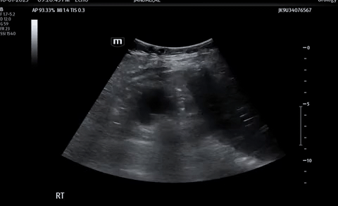
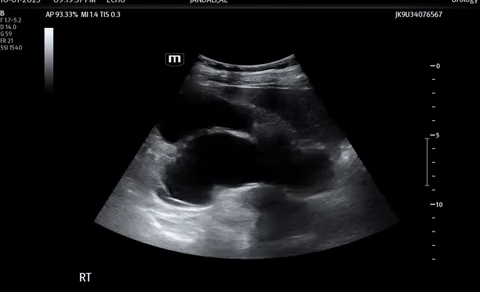
Bedside POCUS
A renal/bladder POCUS was performed to evaluate for any secondary signs of obstruction.
These images demonstrate severe hydronephrosis with obvious loss of renal architecture.
Based on the findings, a CTAP was ordered for further evaluation.
Hydronephrosis
Hydronephrosis is an abnormal enlargement and distension of the kidney due to obstructive uropathy.
It can be bilateral or unilateral and it is critical to evaluate both sides when using ultrasound because it is important for comparison, but also to narrow down the differential diagnosis.
For example, bilateral hydronephrosis may be due to prostate or uterine pathology vs. unilateral hydronephrosis from an obstructing stone.
There are two major grading scales (listed below) however, there is no one universally accepted grading scale.
Mild, Moderate and Severe
Grades I - IV


Both grading scales are essentially on a continuum; subsequent grades have more extensive disruption of the normal renal anatomical structure,, which can thereby affect function.
Earlier grades of hydronephrosis include dilation of the renal pelvis first, then the major and minor calyces become distended and confluent and ultimately, the cortex is no longer preserved and there is effacement of the cortex and medulla.
Renal cysts often mimic hydronephrosis.
However, simple cysts are usually thin walled, without septations, localized to the periphery and do not connect to calyces.
Always scan in both planes to help differentiate the two.
Back to the case:
CTAP showed severe right hydroureteronephrosis secondary to abnormal soft tissue at the UV junction suspicious for urothelial malignancy.
Key Take Home Points
Renal POCUS can help narrow differential diagnoses efficiently and can guide management regarding the necessity of further imaging or consultations
Always evaluate both kidneys for comparison and scan in multiple planes to avoid confusion between hydronephrosis and renal cysts
There are multiple grading scales used to characterize hydronephrosis, but each represent worsening distension of the kidney and ultimate destruction of the normal renal architecture
References
Blok , Barbara K. “Renal Ultrasound.” Www.acep.org, 19 Oct. 2020, www.acep.org/sonoguide/basic/renal-ultrasound.
Bueschen, Anton J. “Flank Pain.” Nih.gov, Butterworths, 2015, www.ncbi.nlm.nih.gov/books/NBK292/.
Dinh, Vi. “Renal Ultrasound Made Easy: Step-By-Step Guide.” POCUS 101, www.pocus101.com/renal-ultrasound-made-easy-step-by-step-guide/.
Koratala, Abhilash. “Cyst or Hydronephrosis?” NephroPOCUS, 3 Aug. 2020, nephropocus.com/2020/08/03/cyst-or-hydronephrosis/. Accessed 9 Oct. 2023.
---. “Hydronephrosis.” NephroPOCUS, 31 May 2019, nephropocus.com/2019/05/31/hydronephrosis/.