
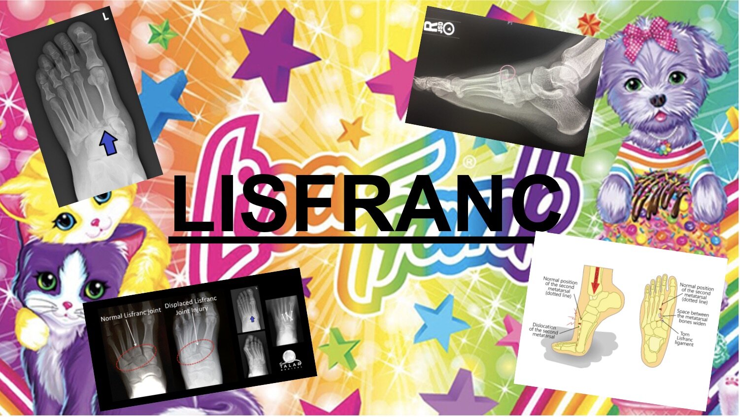
Lisa Frank? No, Lisfranc
Written by Luke Fey PGY-3. Edited by Victor Huang MD
How confident are you that you can properly describe what a Lisfranc injury is? If your answer is not 100%, you should keep reading.
INTRODUCTION
A Lisfranc injury is a tarso-metatarsal fracture dislocation characterized by traumatic disruption between the articulation of the medial cuneiform and base of the second metatarsal. More broadly speaking, Lisfranc injuries refer to any injury (fracture or dislocation) of the Taraso-MetaTarsal (TMT) joint complex. For clarification purposes, I use the term “Lisfranc injury” and “TMT joint injury” interchangeably in this article. The Lisfranc ligament connects the medial cuneiform to the head of the 2nd metatarsal and is a keystone piece for maintaining the “Roman” arch of the midfoot. Disruption of this arch (whether by fracture or ligamentous injury causing subluxation) can lead to foot instability.

A Lisfranc injury is a rare (0.2% of all fractures) but important entity to keep on your differential when working up traumatic foot pain.1 Unfortunately, many of these injuries are missed and can lead to long term morbidity in the form of osteoarthritis, chronic instability, pain, deformity and poor functional outcomes. 20% of these injuries are missed on the first evaluation.2 Therefore, it is essential that the Emergency Physician keep a high index of suspicion for this injury pattern when evaluating patients with traumatic foot pain.

HOW DOES THIS HAPPEN?
The mechanism usually involves rotational and axial forces on a hyper-plantarflexed foot. While high speed motor vehicle accidents and direct crush injuries most commonly cause this injury pattern, it can also occur when falling from a height such as missing a step when going down the stairs. Additionally, this is commonly seen in sports when one player may fall or step on a player who is running in front of them.
PHYSICAL EXAM
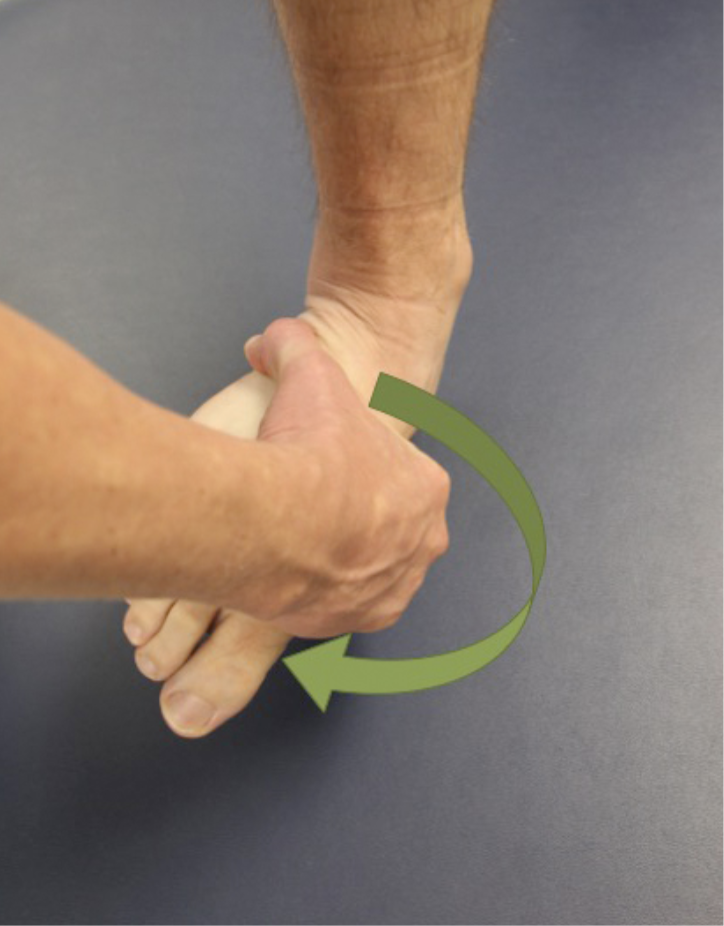
Physical exam findings may be subtle. There may be external signs of trauma (ecchymosis and swelling of the medial mid-foot). Plantar ecchymosis of the midfoot is often considered pathognomonic for a significant TMT joint complex injury (although this finding is rarely present immediately after the injury). Patients will often not be able to bear-weight on their toes. The “piano key” test may exacerbate pain with dorsiflexion and plantarflexion of the affected metatarsal. Pronation and abduction will also particularly exacerbate the pain from this injury.
WORK-UP
AP, lateral and oblique plain films of the affected foot are the first-line test of choice to diagnose this injury pattern. Any obvious fracture to the base of the metatarsals requires immediate referral to a surgical specialist as the majority of these injuries are managed opertively. A pathognomonic finding for Lisfranc injury is the “fleck sign.” This is a bony avulsion at the origin or insertion of the Lisfranc ligament located in the vicinity of the medial border of the head of the 2nd metatarsal.3 A subtle finding is diastasis, or separation, of the space between the 1st and 2nd metatarsal bases.
If no obvious fracture is present, you must look for the following patterns to assure normal anatomic alignment of the bony structures of the midfoot.
Co-linear alignment of the head of the 2nd metatarsal with the medial border of the MIDDLE cuneiform on the AP film
Co-linear alignment of the head of the 4th metatarsal with the medial border of the cuboid on the oblique view
Dorsal and plantar borders of the metatarsals should align with the cuboid and cuneiform bones on the lateral view
***ANY STEP-OFF OR MALALIGNMENT OF THE ABOVE FINDINGS SHOULD WARRANT FURTHER INVESTIGATION***
If plain films fail to demonstrate any abnormality and the patient’s mechanism and physical exam are suggestive of possible injury, weight bearing plain films should be performed. Some studies suggest that even weight bearing films have poor sensitivity for Lisfranc injury and that CT or MRI should be performed on patients with high suspicion for injury.4 CT can definitively rule out any fracture or subluxation of the metatarsals and MRI can rule-in a purely ligamentous injury to the Lisfranc ligament.

ED TREATMENT
This part is easy; ANY injury (fracture or dislocation) to the TMT joint should be immobilized (posterior short leg splint), made non-weight bearing and referred to podiatry or orthopedics. As stated before, these injuries have a high rate of complications and high risk of morbidity so they should be urgently referred to a surgical specialist with experience treating these injuries. There are several classifications of Lisfranc injury that are used to aid in conservative versus operative management in these injuries but they are widely variable and beyond the scope of this article.
For patients with both inconclusive findings on plain film and a CT scan that does not show any subluxation or fracture AND you still suspect a purely ligamentous injury - these patients should be immobilized, made non-weight bearing and referred to a Podiatrist or Orthopedist for an outpatient MRI.
CLOSING REMARKS
Remember the definition of a Lisfranc injury - a fracture dislocation of the head of the 2nd metatarsal as it relates to the medial cuneiform. This is a high-risk injury pattern that should not be missed. Use the history and physical exam to tip you off to possible midfoot instability involving the TMT joint. If there is clinical suspicion despite negative initial plain films, pursue weight bearing films and consider a CT scan. In the end, you are safe to immobilize the extremity, make the patient non-weight bearing and refer all of them to an outpatient surgeon to further investigate their midfoot injury. It will keep them walking in the long run.
REFERENCES
Epidemiology of adult fractures: A review. Court-Brown CM, Caesar B. Injury. 2006;37(8):691. Epub 2006 Jun 30.
Watson TS, Shurnas PS. Treatment of Lisfranc Joint Injury : J Am Acad Orthop Surg. 2010;18(12):718-728.
Fracture dislocations of the tarsometatarsal joints: end results correlated with pathology and treatment. Myerson MS, Fisher RT, Burgess AR, Kenzora JE. Foot Ankle. 1986;6(5):225.
Conventional radiography, CT, and MR imaging in patients with hyperflexion injuries of the foot: diagnostic accuracy in the detection of bony and ligamentous changes. Preidler KW, Peicha G, Lajtai G, Seibert FJ, Fock C, Szolar DM, Raith H. AJR Am J Roentgenol. 1999 Dec;173(6):1673-7.
Welck MJ, Zinchenko R, Rudge B. Lisfranc injuries. Injury. 2015;46(4):536-541. doi:10.1016/j.injury.2014.11.026
Clare MP. Lisfranc injuries. Curr Rev Musculoskelet Med. 2017;10(1):81-85. doi:10.1007/s12178-017-9387-6
DeOrio M, Erickson M, Usuelli FG, Easley M. Lisfranc Injuries in Sport. Foot Ankle Clin. 2009;14(2):169-186. doi:10.1016/j.fcl.2009.03.008
Aziz F, Doty CI. Orthopedic Emergencies. In: Stone C, Humphries RL. eds. CURRENT Diagnosis & Treatment: Emergency Medicine, 8e. McGraw-Hill; Accessed September 24, 2020. https://accessmedicine.mhmedical.com/content.aspx?bookid=2172§ionid=165061678
Omer T, Santiago-Martinez M. Foot Injuries. In: Tintinalli JE, Ma O, Yealy DM, Meckler GD, Stapczynski J, Cline DM, Thomas SH. eds. Tintinalli's Emergency Medicine: A Comprehensive Study Guide, 9e. McGraw-Hill; Accessed September 24, 2020. https://accessmedicine.mhmedical.com/content.aspx?bookid=2353§ionid=222407540